


To scan, or not to scan, that is the question!
Posted on 5th June 2019 at 11:54
We should never discount an MRI scan as it can be used as an extremely accurate method of disease detection throughout the body. However, they can also be quite misleading.
A high proportion of patients that we see come to us with issues concerning their lower back or neck. We are all very experienced and skilled clinicians, and can usually let our patients know what we think their problem is, without scans or x-rays. Many patients though feel that we can only really know what is going on if they have an MRI scan.
MRIs are not indicated clinically if there are no neurological signs and symptoms. These may include pain referred down your arm or leg, pins and needles or numbness. If the pain is confined to the back or neck it may be that an MRI is not indicated.
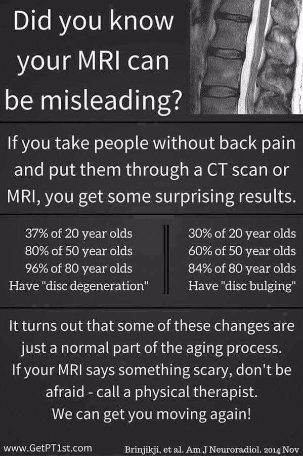
The other issue with MRIs is that they often show problems that are ‘normal’ age related changes. Not all findings are relevant, and the interpretation of those findings is vital. You may be told that you have a disc bulge but unless it is pressing on a nerve it is probably a normal age-related change, and may well not be related to your symptoms. We need to also relate what we see on the scan images with what our patients are presenting with. For example, a scan may show a disc bulge on the left side of the spine, but the symptoms are actually on the right side.
It is very easy to become fearful of what is found on your MRI. We have patients who come in having been told that they have numerous disc bulges and are then worried to bend, lift or move in case they become worse. It takes a long time to unravel that fear avoidance and to encourage people to move again.
We need to be focusing on getting rid of peoples fear, interpreting results correctly and in a manner that is easy to explain to our patients, and getting people to be able to move again, hopefully with much less pain!
Share this post: